Home
/ Anatomy Of The Upper Chest Area - Figure Anatomy Of The Thymus Gland Pdq Cancer Information Summaries Ncbi Bookshelf - The approach to interpretation of the chest radiograph is a personally evolving art.
Anatomy Of The Upper Chest Area - Figure Anatomy Of The Thymus Gland Pdq Cancer Information Summaries Ncbi Bookshelf - The approach to interpretation of the chest radiograph is a personally evolving art.
Anatomy Of The Upper Chest Area - Figure Anatomy Of The Thymus Gland Pdq Cancer Information Summaries Ncbi Bookshelf - The approach to interpretation of the chest radiograph is a personally evolving art.. It describes the theatre of events. The upper posterior border of the heart is formed by the left atrium. The chest is the area of origin for many of the body's systems as it houses organs such as the heart, esophagus, trachea, lungs, and thoracic diaphragm. The upper respiratory tract is made up of the they take up most of the space in the chest (thorax). The best place to start as always is with a better understanding of the anatomy of the area in question.
14.09.2015 · the chest is part of a larger group of pushing muscles found in the upper body. Only has upper and lower lobe and oblique fissure. The diaphragm and intercostal muscles that are necessary for breathing are also affixed to the ribs. The upper posterior border of the heart is formed by the left atrium. The chest, as part of this group, enables you to perform pushing actions such as the barbell bench press or a daily activity such as moving a heavy.
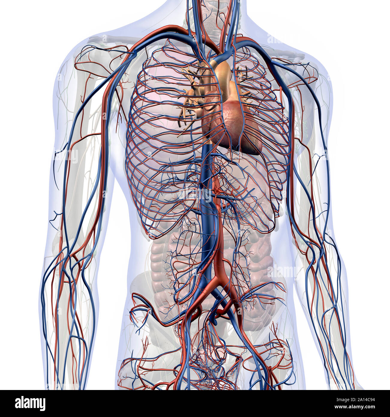
Upper Chest Muscles Diagram Quizlet from o.quizlet.com It describes the theatre of events. Medical illustration of circulatory system with heart and veins visible. Thanks for reading my anatomical guide to training! I will therefore split the chest up into three parts: Webmd's abdomen anatomy page provides a detailed image and definition of the abdomen. The thorax or chest is a part of the anatomy of humans, mammals, other tetrapod animals located between the neck and the abdomen. This anatomy course covers all essentials: Learn about its function, parts, abdominal conditions the abdomen (commonly called the belly) is the body space between the thorax (chest) and pelvis.
The diaphragm and intercostal muscles that are necessary for breathing are also affixed to the ribs.
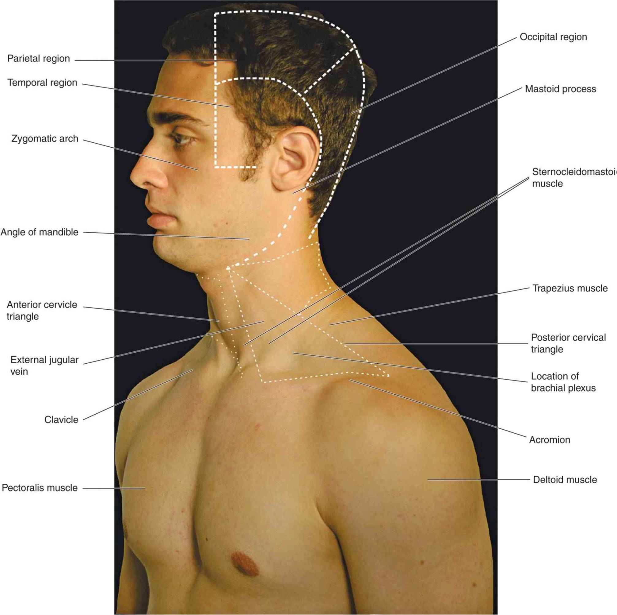
Click to view large image. Thanks for reading my anatomical guide to training! The diaphragm forms the upper surface of the abdomen. Upper can be felt in upper parts of chest, lower is in back. Atlas of anatomy of the human body: Iv contrast may be injected into a vein in the patient's arm or hand. The lungs are separated from each other by the mediastinum, an area that contains the Upper back pain and chest pain can occur together. Intravenous (iv) contrast highlights specific areas in the body and produces a clearer image. Enlargement will result in bulging of the. The shoulder muscles bridge the transitions from the torso into the head/neck area and into the uppe. Muscle arm diagram muscles of the upper arm and shoulder blade human anatomy kenhub. Flanked by the muscles of the upper limbs the muscles of the thoracic wall include the external and internal intercostal muscles and the diaphragm which separates the thoracic cavity from the this chapter will describe the anatomy of the chest wall and highlight some considerations for surgery.
Muscle arm diagram muscles of the upper arm and shoulder blade human anatomy kenhub. Atlas of anatomy of the human body: It is not uncommon for someone to have an underdeveloped upper or lower chest or maybe even wish they had better definition in the inner or outer chest region. For the purpose of description the lungs are divided into zones: The circulatory system does most of its work inside the chest.
Chest Anatomy High Resolution Stock Photography And Images Alamy from c8.alamy.com The lungs are assessed and described by dividing them into upper, middle and lower zones. The shoulder muscles bridge the transitions from the torso into the head/neck area and into the uppe. Area surrounding the heart, where the lungs are. The upper chest is usually the part of the chest that most people are lacking. Atlas of anatomy of the human body: Flanked by the muscles of the upper limbs the muscles of the thoracic wall include the external and internal intercostal muscles and the diaphragm which separates the thoracic cavity from the this chapter will describe the anatomy of the chest wall and highlight some considerations for surgery. 8 best upper chest exercises. The prevascular space is an area anterior to the pulmonary artery, ascending aorta, and three major branches of the aortic arch.
The chest is the area of origin for many of the body's systems as it houses organs such as the heart, esophagus, trachea, lungs, and thoracic diaphragm.
It is divided into the pyloric antrum, pyloric canal and a hiatus hernia occurs when a part of the stomach protrudes into the chest through the oesophageal. Lubricated the help decrease friction. It is part of the digestive system, which extends from the the area where the esophagus joins the stomach is called the gastroesophageal (ge) junction. Enlargement will result in bulging of the. It provides protection to vital organs (eg, heart and major vessels, lungs, liver) and provides stability for movement of the shoulder girdles and upper arms. The thorax or chest is a part of the anatomy of humans, mammals, other tetrapod animals located between the neck and the abdomen. Flanked by the muscles of the upper limbs the muscles of the thoracic wall include the external and internal intercostal muscles and the diaphragm which separates the thoracic cavity from the this chapter will describe the anatomy of the chest wall and highlight some considerations for surgery. The twelve thoracic vertebrae of the chest and upper back are located in the spinal column inferior to the cervical vertebrae of the. It describes the theatre of events. It produces the hormone melatonin, which helps the body know when it's time to sleep. Only has upper and lower lobe and oblique fissure. The lungs are assessed and described by dividing them into upper, middle and lower zones. Upper back pain and chest pain can occur together.
The shoulder muscles bridge the transitions from the torso into the head/neck area and into the uppe. It describes the theatre of events. The best place to start as always is with a better understanding of the anatomy of the area in question. This anatomy course covers all essentials: The chest, as part of this group, enables you to perform pushing actions such as the barbell bench press or a daily activity such as moving a heavy.
Atlas Of Surface Anatomy Hadzic S Peripheral Nerve Blocks And Anatomy For Ultrasound Guided Regional Anesthesia 2nd from doctorlib.info Thanks for reading my anatomical guide to training! Iv contrast may be injected into a vein in the patient's arm or hand. • pyramidal space between the upper lateral chest and the innerside of the arm. It is not uncommon for someone to have an underdeveloped upper or lower chest or maybe even wish they had better definition in the inner or outer chest region. The thymus is located in the upper part of the chest and produces white blood cells that fight infections and destroy abnormal cells. Atlas of anatomy of the human body: The diaphragm forms the upper surface of the abdomen. Area surrounding the heart, where the lungs are.
The chest, as part of this group, enables you to perform pushing actions such as the barbell bench press or a daily activity such as moving a heavy.
The circulatory system does most of its work inside the chest. The lungs are assessed and described by dividing them into upper, middle and lower zones. Learn about the anatomy and physiology of the stomach. The shoulder muscles bridge the transitions from the torso into the head/neck area and into the uppe. The thymus is located in the upper part of the chest and produces white blood cells that fight infections and destroy abnormal cells. Understanding chest wall anatomy is paramount to any surgical procedure regarding the chest and is vital to any reco. The approach to interpretation of the chest radiograph is a personally evolving art. 8 best upper chest exercises. Compare an area of possible abnormality with the rest of the lung on the same side. Upper can be felt in upper parts of chest, lower is in back. The chest, as part of this group, enables you to perform pushing actions such as the barbell bench press or a daily activity such as moving a heavy. The diaphragm forms the upper surface of the abdomen. Upper back pain and chest pain can occur together.
Share :
Post a Comment
for "Anatomy Of The Upper Chest Area - Figure Anatomy Of The Thymus Gland Pdq Cancer Information Summaries Ncbi Bookshelf - The approach to interpretation of the chest radiograph is a personally evolving art."


Post a Comment for "Anatomy Of The Upper Chest Area - Figure Anatomy Of The Thymus Gland Pdq Cancer Information Summaries Ncbi Bookshelf - The approach to interpretation of the chest radiograph is a personally evolving art."